
The universe around us is composed of atoms, and their structures and reactions are the fundamental drivers of how materials and organisms behave. A new facility gives researchers at The University of Texas at Austin the ability to explore their projects all the way to that single atom level.
The new Texas Materials Institute-led Electron Microscopy Facility recently opened in the Cockrell School of Engineering, giving researchers cutting-edge tools for deeper analysis of their projects. The facility represents a world-class upgrade to UT’s electron microscopy capabilities, bringing it to the forefront of nanotechnology research, according to the leaders of the lab.
Understanding atomic structure of matter is an important aspect of building new technology. Changes in atom positioning can lead to dramatic shifts in properties that can only be observed and experimented on with powerful electron microscopy tools. This paves the way for new discoveries and applications across all disciplines of science, engineering and medicine.
“Electron microscopy gives us the tools to see what we have made and then take this information to improve the chemical synthesis and ultimately produce new devices and technology,” said Jamie Warner, director of the new facility and professor in the Walker Department of Mechanical Engineering and the Department of Electrical and Computer Engineering. “This can lead to improvements in energy storage materials, developing new quantum materials, understanding the behavior of exotic semiconducting and superconducting materials and watching chemical reactions as they happen with atomic-level vision.”
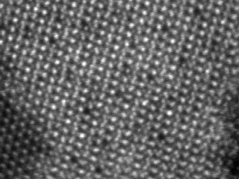
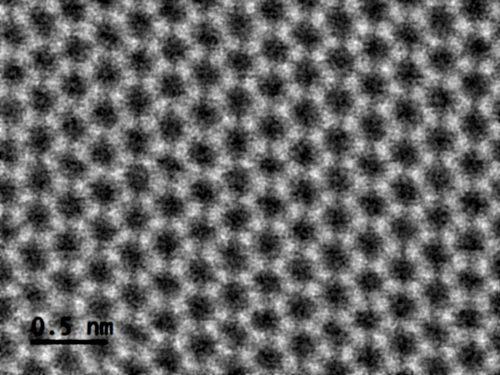
Examples of results using the facility's high-powered microscopes
The facility stands out from others because of its unique capabilities to study fragile samples that would decompose in air or under the harsh conditions of the electron imaging process, using low-energy electrons and cryogenic sample preparation and imaging methods. A special, ultrafast pixelated detector can record single electrons after they interact with a sample to create atomic-scale images with much lower doses and higher energy than conventional systems, Warner said. These techniques will enhance the ability to create new, ultra-thin 2D materials and soft materials, including polymers and organics.
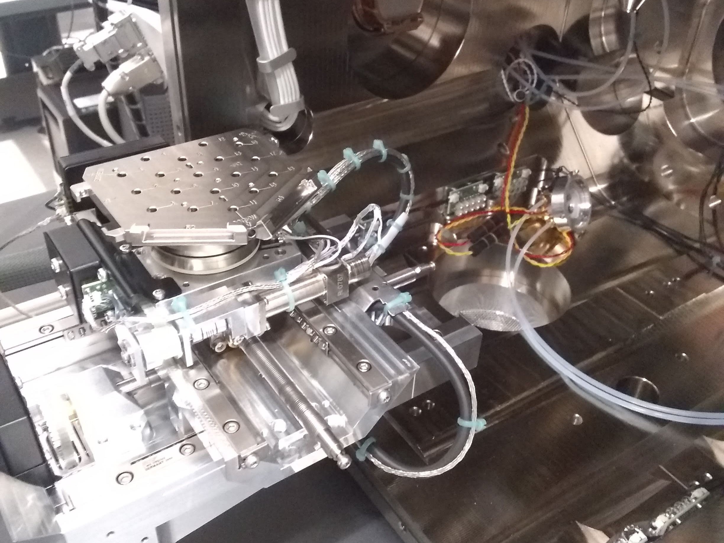
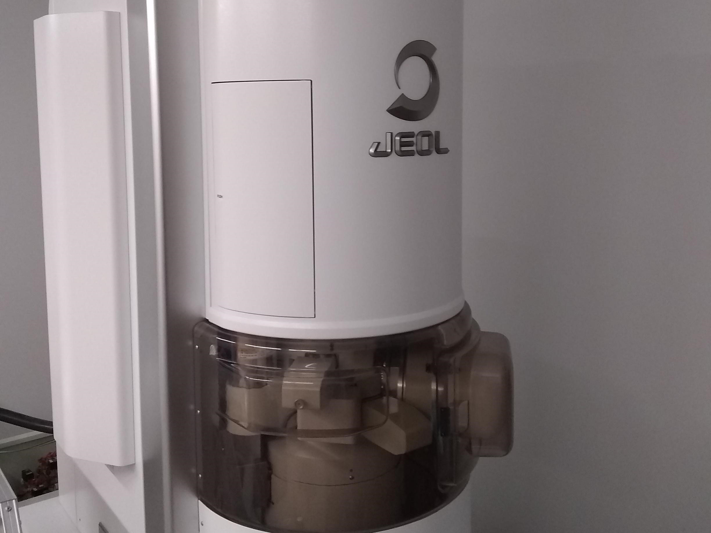
The centerpiece of the facility is a JEOL neoARM Scanning Transmission Electron Microscope (STEM), which offers the ability to analyze samples at the atomic level. A Focused Ion Beam Scanning Electron Microscope (FIB-SEM) uses gallium ions to cut up materials to create a cross-section for observation and analysis. A second SEM provides additional imaging capabilities.
The microscopes are capable of advanced imaging techniques such as 4D STEM and cryo-imaging that can help researchers see things normal cameras can’t capture. One example of a project using these technologies involves freezing batteries, slicing them up using the FIB-SEM and analyzing the samples to learn more about why the batteries failed.
The $10 million facility is located on the ground floor of the Cockrell School's Engineering Education and Research Center and will operate under the Texas Materials Institute, UT’s materials science and engineering research center and graduate program.
Warner joined the Cockrell School in January after spending 13 years at Oxford University’s Department of Materials, where he led the Nanostructured Materials Group.
“Having worked with some of the top electron microscopy tech in the world at Oxford, I’m excited to see UT’s commitment to building top-notch facilities and becoming a world leader,” Warner said.
With the ongoing COVID-19 pandemic, classes and trainings at the facility have been postponed. A minimal onsite presence remains, and critical experiments, such as those needed for graduation or grant deadlines, receive priority.
For more information, visit the Electron Microscopy Facility website.

